Archive for April, 2013

Swollen Fingers Causes, Symptoms and Treatment

Apr 30 2013 Published by under Diseases & Conditions

What are Swollen Fingers?

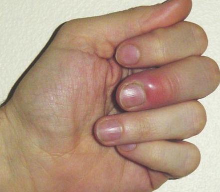
A swollen finger is a medical sign of inflammation or fluid build-up in the joints or tissues that are located in a person’s finger. The fingers are one of the common body parts that tend to be overused. Hence, they are not given significant attention unless they become swollen. When a person has swollen fingers, he or she will not be able to perform their normal or day-to-day activities like holding, eating, and touching.

Swollen finger

Swollen Fingers Causes

The etiological factors that cause swollen fingers are as follows:

  • Autoimmune, degenerative, inflammatory, and infectious etiological factors include:
    • Carpal tunnel syndrome
    • Infections like streptococcus aureus infection
    • Osteoarthritis
    • Septic arthritis
    • Rheumatoid arthritis
    • Paronychia
    • Ganglion cyst
    • Cellulitis (bacterial skin infection)
    • Bursitis (inflammation of the bursa)
    • Herpetic withlow
  • Injury etiological factors include:
    • Repetitive stress injury
    • Cartilage or ligament injury
    • Broken bones
    • Blunt or laceration of the bone

When one has one of the etiological factors mentioned above, he or she will experience the condition called swollen fingers. The complications that are associated with swollen finger condition include finger deformity, surgical repair damage, spread of infection to nearby areas, inability o perform normal day-to-day tasks, finger amputation, and chronic form of disability.

Swollen Fingers Associated Symptoms

When a person has a swollen finger, he or she may have an underlying disease condition that needs to be checked immediately. Such condition includes infections which may be accompanied by chills, redness, warmth, and fever. Other associated symptoms that may be experienced include the following:

  • Fatigue
  • Bluish discoloration
  • Pain
  • Stiffness
  • Decreased movement or motion
  • Pus-filled cracks
  • Laceration
  • Lumps on fingers
  • Swelling of the joints

Swollen Fingers Treatment

There are two suggested treatments for a person with swollen fingers. The first one uses the acronym RICE, and the other is a home remedy treatment.

RICE

The medically accepted acronym RICE stands for:

  • Rest
  • Ice
  • Compression
  • Elevation

First, the affected finger needs to rest, and ice compression is applied for at least ten minutes to the swollen area. Next, a bandage or tape is applied to cover and wrap the swollen finger but not too tightly to allow blood circulation. Lastly, the affected area needs to be elevated, e.g., to have it in an upright position. This method should be repeated until the swollen area gets better.

Home Remedies

Another treatment option for swollen fingers is home remedy treatment which includes the following:

Garlic

Garlic has a substance that is called allicin, which is known to assist a person’s immune system wherein it suppresses microorganism reproduction and growth. In the home remedy treatment, 2-3 gloves of garlic are combined with mustard oil in a small bowl, and the mixture is heated. Once the heated mixture has cooled down, the oil is applied to the swollen finger and left on the finger for an hour or so. The procedure is repeated at least 4-5 times to get instant relief.

Turmeric

Turmeric is known to be a natural kind of antiseptic herb powder with healing properties. Its function is to kill bacteria that causes pain and inflammation. In the home remedy treatment, turmeric powder and a bit of water are mixed create a paste form which is applied to the swollen finger. The affected finger is then wrapped with cotton. After two hours of application, the affected area is to be washed with lukewarm water.

Tea tree oil and olive oil

Tea tree oil has antibacterial, antiviral, and antifungal properties, while olive oil has a soothing effect on the skin. Hence, the mixture of these two components will be an effective treatment for swollen fingers. In the home remedy treatment, small equal amounts of both oils are mixed together and heated. Afterwards, the mixture is applied to the swollen finger. For best results, it is advised to treat the finger before bedtime and to leave the mixture on overnight.

Epsom salt

This is another component that will treat swollen fingers. The Epsom salt is effective when the etiological factor of the swollen fingers is an electrolyte imbalance. Epsom has sulfate and magnesium, and the skin would be able to absorb these elements without any complications. In the home remedy treatment, the swollen finger is soaked in a mixture of Epsom salt and warm water for 10-15 minutes or so. To achieve desired results, this method is to be repeated daily. In cases when Epsom salt is not available, normal salt, which provides the same effect, can be used instead.

These are the common components of home remedy treatments that can be used to treat swollen fingers. The suggested mixtures or preparation and applications will help heal swollen fingers.

No responses yet

Internuclear Ophthalmoplegia

Apr 28 2013 Published by under Eyes & Vision,Uncategorized

What is Internuclear Ophthalmoplegia?

Internuclear ophthalmoplegia (INO) is a condition that affects the conjugate lateral gaze, resulting in problems in eye adduction. The condition involves the impairment of horizontal eye gaze due to disruptions in the connection between cranial nerve centers within the brain. The affected eye usually cannot turn inwards but can turn outwards during horizontal gaze.

The exact phenomenon involves a minimal adduction of the contralateral eye when a person attempts to see something from the contralateral side. The unaffected eye abducts with associated nystagmus. In simpler terms, when the eyes gaze towards one side, the affected eye does not turn to the side to look at the object, but rather it stays fixed in the middle, while the unaffected eye is able to turn to the side to look at the object but undergoes involuntary rapid movement called nystgamus.

The eyes may also have horizontal diplopia which involves double vision in the affected eye when it looks towards the opposite direction (for example, when the right eye is affected, it suffers double vision when it looks towards the left).

Internuclear Ophthalmoplegia Symptoms

Symptoms of INO often include:

  • Painless onset of disturbance in vision
  • Absence of diplopia in primary gaze
  • Presence of horizontal diplopia in lateral gaze
  • Problems with adduction on the affected eye
  • Nystagmus on the unaffected eye with abduction

Most commonly, INO is unilateral, but some people may experience bilateral INO. Bilateral affectation may involve medial rectus palsy. Patients may also have no problems in convergence in posterior INO and bilateral INO, but some people with anterior INO may experience problems in convergence.

Causes

Causes of INO include dysfunction in the medial longitudinal fasciculus (MLF), which is a heavily myelinated tract that connects the oculomotor nucleus to the paramedian pontine reticular formation. This mechanism allows conjugate movements of the eye. INO can also be caused by other factors such as:

Multiple sclerosis

MS causes bilateral INO in young patients due to demyelination of the nerves. INO in young patients is usually bilateral as a result of multiple sclerosis.

Stroke

Stroke also causes unilateral INO, affecting the ipsilateral side of the brain. Stroke may be in the form of brainstem infarction or other related types. Stroke is the most common cause of INO in older patients.

Myasthenia gravis

Myasthenia gravis (MG) can produce a pseudo-INO.

Others

  • Viral infection
  • Fourth ventricular tumors
  • Syphilis
  • Trauma
  • Lyme disease
  • Subdural hematoma
  • Drug intoxication from tricyclic antidepressants and phenothiazine

Pathophysiology

The presence of synchronous eye movements is the responsibility of several structures in the eye and brain. The cranial nerves VI, IV, and III communicate through the medial longitudinal fasciculus (MLF). The medical nerve fasciculus is the main pathway for nerve impulses from the cranial nerves to produce normal eye movements. In INO, there is possibly lesion in the pathway, disrupting the communication between the cranial nerve nuclei and the MLF.

For instance, in order to look at the right side, the control center of the left horizontal eye movement must send signals to the left cranial nerve VI nucleus, while that of the right horizontal eye movement must send signals to the right cranial nerve VI nucleus. However, when lesions occur in the left and right pathways, the cranial nerves may not be able to send signals to the medial rectus, thereby causing problems in gaze. Due to this, the unaffected eye assumes a lateral gaze through abduction, while the affected eye will not adduct to follow the gaze. The left eye will also go into abducting nystagmus.

Certain infections may damage the neural pathway, thereby leading to INO.

Diagnosis

Diagnosis of INO involves identifying possible causes of the condition in order to come up with the proper treatment regimen. Diagnostic tests include:

  • MRI of the brainstem

MRI of the brain stem may identify lesions in the area that may cause INO.

  • VDRL

Venereal disease research lab (VDROL) testing is a special diagnostic test to identify the presence of syphilis. This involves blood tests to check antigens and antibodies in the patient.

  • Lyme titer

Lyme titer is done to check the presence of Lyme’s disease, which is a condition cause by deer ticks.

  • Fasting blood glucose determination

Fasting blood glucose is especially performed on patients with drug intoxication because drug intoxication may lead to lowered blood glucose levels.

  • Blood pressure measurement

Blood pressure measurements are also taken to monitor post-stroke patients in order to prevent another brain attack episode that may result in further affectation of the cranial nerves.

Treatments of Internuclear Ophthalmoplegia

Treatment for INO involves managing the root cause in order to improve the communication of the cranial nerves and the medial rectus fasciculus. Treatments may include:

Surgery

Surgery may be done to remove brainstem lesions that are causing INO. Surgery is carefully performed as the brain stem is a delicate tissue. Surgery may employ resection of the medial rectus and lateral rectus.

Antibiotic and antiviral therapies

Conditions caused by infections such as viral diseases, Lyme disease, and syphilis are usually treated with medications to kill the causative microorganism.

Steroids

Corticosteroids may be used to reduce cerebral edema and increased intracranial pressure (ICP) in cases of ICP due to a cerebrovascular accident and subdural hematoma.

These treatments regimes specifically correct the underlying problem, which eventually result in correcting the internuclear opthalmoplegia.

Complications

Complications of INO is often a result of complications of various causes. Brain stem lesions may result in respiratory problems. The INO, which involves problems in horizontal gaze, may cause loss of self-esteem due to the visual defects that are apparent to other people. They may also withdraw from socialization due to INO. In terms of learning, there may be no problems in cognitive learning, but vision problems may cause the person to have difficulty in school activities.

No responses yet

Cervicogenic Headache – Symptoms, Treatment, Causes, Exercises

Apr 25 2013 Published by under Diseases & Conditions

What is Cervicogenic Headache?

Cervicogenic headache is a type of headache which involves hemicranial pain that tends to be chronic. The pain is often referred to the head from any soft tissues or bony structures on the neck. The mechanism of cervicogenic headache is rooted from the functional convergence of the trigeminal sensory pathway and the upper cervical pathways. The trigeminocervical nucleus in the upper cervical spinal cord sends pain signals to the sensory fibers in the upper cervical roots. As a result, there is referred pain from the neck to the face and head.

 Cervicogenic Headache

Cervicogenic headache has a different mechanism from other types of headache, but it can be similar in presentation to hemicranias continua, and tension-type and migraine headaches.

The presence of cervical muscle soreness and neck pain is common in primary headache types such as migraine and tension-type headaches. However, in the case of cervicogenic headache, the pain actually arises from the bony and soft structures of the neck, contrary to primary headaches where the neck pain is secondary to the headache. The exact roots of the headache are from problems in the vascular, articular, osseus, neurogenic, or muscular structures of the neck innervated by C1 to C3 cervical vertebrae.

The presence of cervicogenic headache is often initiated by awkward head position or neck movements.

Cervicogenic Headache Symptoms

Symptoms of cervicogenic headache include:

Ipsilateral headache

A headache is the most apparent symptom of cervicogenic headache. The headache usually arises on the same side where neck or arm discomforts are felt. The presence of pain on the occipital area or back of the head usually radiates to the forehead or temples. The quality of pain may be different from person to person. Others may experience an ice pick headache, while others may feel dull pain. The pain in the head usually starts at the base of the neck and progresses up to the head.

Restricted cervical range of motion

The main problem in cervicogenic headache can be traced to neck problems. Patients who suffer from a cervicogenic headache usually experience restricted motions of the cervical spine or neck stiffness. The base of the neck is often tender at the onset of headaches.

Arm, shoulder, and neck pain that tend to be ipsilateral

The discomfort also radiates to the arms and shoulders that occur on the same side of the headache. Patients who suffer from a migraine headache may also experience these discomforts, making the identification of cervicogenic headache difficult.

Sensitivity to light and sound

Similar to a migraine, the cervicogenic headache may also cause increased sensitivity to light and sound, which usually triggers neck and head discomforts.

Nausea and Vomiting

Nausea and vomiting are also classic signs of headaches due to the affectation of nerve roots in the brain stem.

Causes of Cervicogenic Headache

The main causes of cervicogenic headache are neck injuries which cause compression of the occipital nerves. There are various causes of headache including:

  • Trauma

Trauma to the spine such as whiplash injury and other neck-related injuries may cause injury to the neck structures, thereby leading to compression of the cervical nerves and trigeminal nerve.

  • Cervical osteoarthritis

The presence of joint degeneration in the cervical spine may also cause cervicogenic headache due to disruption in the nerve fibers as a result of nerve compression in the area.

  • Tumor growth

Tumors that grow in the spine may also cause occlusion or compression of the adjacent nerves, thereby leading to cervicogenic headache.

  • Frequent downward positioning of the head

Frequent reading or working with the head in a downward position puts strain on the neck muscles and nerves.

  • Improper sleeping and work position

Chronic postural strain from poor working and sleeping positions creates repetitive stress on the neck area, leading to nerve irritation and vascular constriction.

  • Inflammatory condition involving the neck and head

Presence of other inflammatory conditions in the neck may also predispose the occurrence of cervicogenic headaches.

  • Conditions such as diabetes and vasculitis

The presence of these disorders usually affects the circulation in the body, including the neck. Vasculitis and diabetes usually result in affectations of the nerves due to poor blood supply in the area.

These causes of cervicogenic headache should be identified in order to differentiate cervicogenic headache from other primary headache types.

Treatment of Cervicogenic Headache

The treatments for cervicogenic headache often employ various modalities such as medications, physical therapy, psychological and behavioral therapies, anesthetic use, and surgery.

Pharmacologic therapy

Medications are used to relieve symptoms of the disorder. These may include:

Analgesics

Centrally acting analgesics as well as non-steroidal anti-inflammatory drugs are given to relieve pain in the neck, head, shoulders,and arms. Narcotic analgesics are rarely used for the treatment of chronic conditions due to the possibility of drug dependence. Analgesics may include acetaminophen, celecoxib, and naproxen.

Antidepressants

Antidepressants are also used for the management of neuropathic pain. Serotonin and norepinephrine reuptake inhibitors such as duloxetine and venlafaxine are most commonly used. The dosage of antidepressants for pain management is significantly lower than that for depression management.

Anticonvulsant drugs

Anticonvulsant drugs are also used for the treatment of neuropathic pain by stabilizing the nerve impulse transmission. These include divalproex sodium, gabapentin, topiramate, and carbamazepine. Liver enzyme should be monitored because these medications usually are hepatotoxic.

Muscle relaxant

Muscle relaxants may also be used to treat cervicogenic headache by relaxing muscles in the neck area, thereby reducing neck stiffness. These may include baclofen and tizanidine hydrochloride.

Botulinum toxin injections

These may be employed to reliefve muscle spasms in the neck.

Steroid injections

Steroids are also employed to reduce inflammation and pain in inflammatory disorders in the neck.

Physical Therapy

Physical therapy serves as a rehabilitative treatment for cervicogenic headache patients. Exercise programs geared in the neck and shoulder area help patients regain normal neck functions.

Psychological and behavioral therapies

These therapies, which serve as adjunctive therapies to allow increased tolerance to pain, use biofeedback, cognitive-behavioral therapy, and relaxation techniques.

Anesthetics

Anesthetics are sometimes injected in the cervical epidural area to provide temporary pain relief. The anesthesia is usually injected in the C2 and C3 levels.

Surgery

Surgery serves as a last resort for treatment of unresponsive patients. Surgical procedures may include the removal of cervical entrapment of the occipital nerve in the trapezius muscle.

ICD 9 code

Cervicogenic headache has an ICD 9 code of 307.81.

No responses yet

Erythema Toxicum Neonatorum

Apr 21 2013 Published by under Diseases & Conditions

What is Erythema Toxicum Neonatorum?

Erythema toxicum neonatorum (ETN), also known as erythema toxicum and toxic erythema of the newborn, is a very common skin lesion that appear on a newborn infant in the first week of life. It was first described by Barthalomaeus Netlinger in 1472 and renamed by Leiner as Erythema neonatorum toxicum in 1912.

Lesions are present not at birth but usually within 12 to 48 hours after birth, and a delayed onset of ETN is sometimes also observed. The lesions, which are benign, may be found scattered anywhere on the body but more commonly in the face and trunk. ETN is a self-limited eruption that resolve spontaneously. It occurs approximately 50 to 70 percent of all full-term newborns but rarely in premature infants or in babies weighing less than 2.5 kilograms. There is no racial or sexual predilection, but there is an increased frequency among infants with increased birth weight and gestational age. Recurrence has been reported during the first two weeks.

Diagnostic exams which involve microscopic examination using a Gram stain or Wright stain will reveal predominance of eosinophils. A smear of central vesiculopustular lesion stained by Wright stain demonstrates numerous eosinophils.

Tsanck smear or fluorescent antibody testing consists of removing the blister roof and preparing a slide from scrapings of the base of the blister using a scalpel blade. This is to determine if it is herpes simplex or ETN.

Skin biopsy may sometimes be needed to rule out other more serious diseases. Lesional skin biopsy obtained for routine histopathology examination reveals subcorneal intraepidermal blistering with the blister cavity filled predominantly with eosinophils. Bacterial cultures of these pustules invariably result in negative findings.

Differential diagnosis includes Herpes simplex virus infection, impetigo, listeria infection, neonatal sepsis, and varicella.

Symptoms of Erythema Toxicum

Lesions

There are four kinds of localized lesions: macules, wheals, papules, and pustules. They first appeared on the face and then spread to the trunk, proximal extremities, and buttocks. The lesions are generally characterized as a rash, usually small (less than 3mm.), pale yellow papules which may be surrounded by erythematous wheal. The lesion appearance is similar to a “flea bite.” Central vesicle may contain numerous eosinophil, and peripheral blood contains eosinophilia up to 20 percent.

Histologically, the pustules are most often located in the epidermis just beneath the stratum corneum and are filled with eosinophils. Histological examination of a macule usually shows a perivascular infiltrate composed primarily of eosinophils and a few polymorphonuclear leucocytes and mononuclear cells.

Erythema toxicum can be confused with herpes simplex infection, impetigo, or miliaria rubra (heat rash). Herpes simplex lesions, which is usually caused by the vertical transmission from mother to newborn, tend to have a more vesicular appearance rather than pustular. Impetigo usually has more developed pustular lesions and can be differentiated based on Gram stain performed on intralesional contents. Presence of neutrophils on Wright stain or presence of organisms on Grain stain is suggestive of bacterial impetigo. Diagnosis of miliaria rubra typically follows from a history of excessive warming, either from occlusive clothing or from an incubator. Lesions are usually more confluent than those seen in erythema toxicum and demonstrate lymphocytic infiltrate on Wright stain.

Usually these lesions are temporary, for they appear during the first or second day after birth and disappear after one week. Full-term newborns who weigh greater than 2.5 kilograms are more likely to show rashes. New lesions appear as old ones resolve.

Causes of Erythema Toxicum

The medical condition has been considered idiopathic for the causes of erythema toxicum have not yet been established. Presently, there are several unproven etiological hypotheses, including skin reaction to absorbed enterotoxines, hence the name “toxicum,” an allergic reaction due to eosinophilia in the lesions (and in the peripheral blood of some patients); however, an allergic agent (e.g., detergent soap) or allergens in bedding and clothing have not yet been identified. Transient adjustment of the newborn’s skin to mechanical/thermal stimuli, a decrease in corticosteroid blood levels following neonatal stress enhancing the eosinophilic response, maternofetal transfer of lymphocytes prior to, or during, delivery causing a minor transient form of acute GvH reaction, and activation of skin antimicrobial system before birth and strengthened in the first few days of life, all have also postulated as factors causing this acute inflammatory response.

Treatment

Because erythema toxicum is a self-limiting skin eruption, no specific treatment is required. When the eruption, which resemble erythema toxicum, presents in a newborn, it is necessary to consider the differential diagnosis and to rule out other potentially serious vesiculopustular dermatoses of neonates that can mimic erythema toxicum clinically.

Complications

There are no reported complications of erythema toxicum. It is only transient and benign, and it remains for a brief duration and resolves spontaneously. Post-inflammatory hyperpigmentation may follow but resolves within weeks to months.

Erythema Neonatorum Pictures

Erythema Toxicum Neonatorum images

Erythema Toxicum Neonatorum

Erythema Toxicum Neonatorum

No responses yet

Addisonian Crisis

Apr 18 2013 Published by under Endocrine System

Definition of Addisonian Crisis

Addisonian crisis is another term for acute adrenocortical insufficiency, hypoadrenalism, and hypocortisolism. Based on statistics, Addisonian crisis is a rare disease and occurs in both women and men alike. It is a disease that affects the adrenal glands and leads to the underproduction of steroid hormones. It is categorized into two types: primary and secondary adrenal insufficiency.

Because this kind of disease condition progresses slowly, the symptoms are usually ignored until they reach a severe form of the disease condition which leads to life-threatening and overwhelming complications.

Other experts would define it as a life-threatening disease condition that occurs when a person has insufficient amount of cortisol, a hormone that is being produced by the adrenal glands. Moreover, people who suffer from Addisonian crisis experience chronic deficiencies of mineralocorticoid and glucocorticoid.

Addisonian Crisis Signs and Symptoms

People who experience Addisonian crisis will report the following symptoms and signs:

  • Fever
  • Confusion
  • Weakness
  • Hypoglycemia
  • Hyperkalemia
  • Tachycardia
  • Hyponatremia
  • Dehydration
  • Hypotension
  • Unintentional weight loss
  • Chills
  • Nausea
  • Loss of consciousness
  • Headache
  • Flank pain
  • Anorexia
  • Chronic constipation
  • Dizziness
  • Vertigo
  • Joint pain
  • Excessive and unusual facial or palmar sweating
  • Slow movement
  • Vomiting
  • Abdominal pain
  • Skin hyperpigmentation
  • Salt craving
  • Diarrhea
  • Irritability
  • Decreased appetite
  • Depression

Addisonian Crisis Causes

Researchers have grouped the etiological factors of Addisonian crisis into categorical groups. For example:

  • Impaired steroidogenesis

Scientifically, cortisol is formed by the adrenal glands. To form cortisol, cholesterol is needed to be biochemically converted into hormones, particularly steroid hormones. When the cholesterol production is interrupted, cortisol is not produced, leading to impaired steroidogenesis.

  • Adrenal dysgenesis

This is another cause of Addisonian crisis. This categorical group type is genetic in origin, and such causes are rare. The gene responsible for this kind of the disease includes DAX-1 gene, ACTH receptor gene, and SF1 transcription factor.

  • Destruction of the adrenal

This is a known etiological reason for people who have Addison’s disease. The destruction of the adrenal cortex is caused by an immune reaction.

  • Corticosteroid withdrawal

When a person uses high dosages of steroid for one or more weeks, he or she will likely produce an adrenal gland suppression due to the presence of exogenous glucocorticoids which is known to suppress the hypothalamic corticotrophin-releasing hormone and pituitary adrenocorticotropic hormone (ACTH). When there is prolonged suppression of the adrenal glands, its atrophy occurs, and the adrenal glands may need more time to reach their recuperation stage.

An impaired organ or medication withdrawal can lead to Addisonian crisis, so it is very important for people to be informed about the disease.

Addisonian Crisis Treatment

The treatment for Addisonian crisis involves hormonal replacement treatment which is used to correct the steroid hormonal level. The treatment options include the following:

Pharmacological treatment

Androgen replacement treatment

People with Addisonian crisis need androgen replacement treatment such as dehydroepiandrosterone. Furthermore, it improves sexual and libido satisfaction.

Corticosteroid injections

This will treat the low corticosteroid level found in people with Addisonian crisis.

Oral corticosteroids

The physician may prescribe fludrocortisones to replace aldosterone. Other medications include prednisone, cortisone acetate, and hydrocortisone.

Antibiotic treatment

This may be given if the cause of the Addisonian crisis is due to infection.

Other medications include the following:

    • Hydrocortisone
    • Dextrose
    • Saline solution

These are the common treatments given to people with Addisonian crisis. The treatments are basically symptomatic in approach.

Risks of Addisonian Crisis

The following are the risk factors associated with Addisonian crisis:

  • Trauma
  • Dehydration
  • Surgical procedure
  • Infection
  • Physical stress
  • pituitary gland or adrenal injury
  • Termination of steroid treatments
  • Cancer
  • Family medical history of Addison’s disease
  • Tuberculosis
  • Thyroid disease
  • Diabetes

Those who are at risk of Addisonian crisis should be informed about this condition, the treatment of this disease, and its prevention.

No responses yet

Discoid Lupus Erythematosus – Pictures, Treatment, Symptoms

Apr 15 2013 Published by under Diseases & Conditions

What is Discoid Lupus Erythematosus?

Discoid lupus erythematosus (DLE) is a medical condition that affects the person’s skin. It is also categorized as an autoimmune and chronic kind of disorder which is common in women. People with DLE, when exposed to the sun, have exacerbated symptoms.

According to studies, people who have DLE will later develop systemic lupus erythematosus (SLE). Compared to SLE, DLE is less serious and can be divided into three categories: generalized, childhood, and localized DLE. Among the three categories, the generalized type is the one that is less common. When a person has localized DLE, he or she will manifest skin lesions. Childhood DLE will produce the same symptoms as those found in adults with DLE.

Discoid Lupus Erythematosus Symptoms

People suffering from DLE have usually reported to have skin rashes and inflammation. The symptoms associated with DLE vary from one person to another, and they can occur occasionally or daily. The symptoms linked to DLE include the folllowing:

  • Scars from lesion which may become permanent
  • Chest pain
  • Pale lips
  • Sun sensitivity
  • Palpitation or rapid heart rate
  • Irregular heart rate
  • Red plaques that appear on the skin, particularly on the forehead, ears, nose, and cheeks
  • Red patches on skin which are crusty or scaly in nature
  • Hair loss
  • Episodes of breathing difficulty
  • Itchy feeling
  • Wart-like lesions
  • Surface scaling due to keratin
  • Arthalgia (pain in joints)
  • Blocked follicles due to keratin buildup
  • Development of new lupus plaques
  • Reticulate telangiectasia (skin blotching), particularly in the leg region
  • Chilblains
  • Raynaud’s phenomenon

When a person experiences one or some of the mentioned symptoms, it is highly recommended to consult a physician receive proper treatment.

Discoid Lupus Erythematosus Causes

Currently, the cause of why DLE is being experienced by some is still unknown or idiopathic in nature. The only known information about DLE is that it is an autoimmune kind of disorder, i.e., the person’s immune system has erroneously attack his or her own cells , leading to its destruction and to the person’s illness. Other research studies have proven that gene mutation plays a role in DLE.

Discoid Lupus Erythematosus Treatment

It is important to emphasize that DLE has no cure. Treatment management that is mentioned below is given to control the symptoms associated with DLE until the disease condition can improve on its own, usually attained after a person has undergone treatment management for a few months or even years. The following treatments are suggested for people who suffer from DLE:

Systemic therapy treatment

Here a person with DLE is given a course of oral corticosteroids  in systemic doses.

Topical therapy treatment

Under this therapy, people with DLE are advised to:

  • apply sunscreen to sun-exposed skin areas
  • apply corticosteroid ointment to affected skin areas

Laser and excision therapy treatment

People with DLE can also opt for laser and excision therapy which is done to remove scarred and burned-out lesions.

Other treatment options include

    • Antimalarial drugs
    • Psoriasis medications
    • DMARDs (disease modifying anti rheumatic) drugs
    • Plastic wrapping of the skin after steroid application

It should be stressed that people with DLE should avoid excessive sun exposure as much as possible since it is known to exacerbate DLE symptoms. They can do so by applying sunscreen even on cold or cloudy days. Furthermore, it is advisable to use sun-protective equipment such as hats, umbrellas and the like and to protect their skin at all times especially when they are outdoors. Moreover, it is recommended to use fluorescent lights that emit less or no ultraviolet radiation.

Long-term medication treatment

The long-term medication treatment needs follow-up check-ups which are done at regular intervallic periods. People with DLE will undergo routine laboratory exams such as urinalysis, complete blood count, and renal function tests. These are necessary to check the DLE disease condition.

Pictures of Discoid Lupus Erythematosus

Discoid Lupus Erythematosus
Discoid Lupus Erythematosus picture
Discoid Lupus Erythematosus pictures

No responses yet

Corneal Abrasion – Treatment, Symptoms, Causes, Diagnosis

Apr 10 2013 Published by under Diseases & Conditions

What is Corneal Abrasion?

Corneal abrasion is the medical condition that involves the loss of the epithelial surface layer of the eye’s cornea. The corneal abrasion, which can be either large or small, is a common kind of eye injury that is sometimes neglected. This is due to a disruption of the corneal epithelium integrity or the fact that the surface of the cornea has been denuded or scraped away as a result of external physical forces.

On the one hand, other experts define corneal abrasion as a scratched, torn, or scraped corneal epithelial layer (the surface area of the cornea) which is caused by injury from protruding objects, damage from contact lens or flying glass. On the other hand, others would define corneal abrasion as a loss of tissue from the corneal epithelium which can be either deep or superficial due to trauma to the eye. It comprises a defect in that area with or without breach of Bowman’s layer.

Corneal Abrasion

Corneal Abrasion Symptoms

People who have recently suffered an eye injury are susceptible to corneal abrasion. The following are symptoms which may be experienced by people who suffer from corneal abrasion:

  • Eye pain (especially when the eye is exposed to bright light)
  • Teary eyes
  • Distortion or blurring of vision
  • Spasm in the eye muscle
  • Excessive squinting
  • Eye redness
  • Inability to open the affected eye
  • Mild photophobia
  • Foreign body sensation
  • Edema in the epithelial area
  • Swollen eyelids
  • Mild anterior chamber reaction
  • Crust build-up
  • Headache

People with corneal abrasion are often unaware that they have this condition. Usually the most common symptoms associated with corneal abrasion can be felt and seen after the eye injury.

Corneal Abrasion Causes

People  may experience corneal abrasion due to the following causes or reasons:

  • Corneal injury
  • Walking into a tree branch
  • Corneal dystrophy like lattice corneal dystrophy
  • Trapped foreign object under the upper eyelid
  • Penetration of projectile particles
  • Finger poking
  • Excessive eye rubbing
  • Hot cigarette ash in eye
  • Exposure to sunlight or ultraviolet light
  • Eye infection
  • Object blown into, or poked in, the eye
  • Sport injuries
  • People who have undergone surgical procedure that required general anesthesia
  • Over-prolonged contact lens wear

Corneal Abrasion Diagnosis

People who are suspected to have corneal abrasion are usually recommended to undergo tests like:

  • Physical examination
  • Medical historical examination
  • Slit lamp examination
  • Visual acuity examination
  • Fluorescein staining examination (confirmative diagnostic test)
  • Orbit CT scan

Corneal Abrasion Treatment

Generally a person who suffers from a small corneal abrasion requires no treatment. However, large abrasions require treatment. Minor abrasion or scratches will generally heal in two days. Treatment for corneal abrasion includes:

Medical treatment

The medications usually prescribed for people who suffer from corneal abrasion include analgesics and antibiotics which can be through ointment or eye drops. Normally, corneal abrasion heals rapidly. Hence, there is a need to manage the corneal abrasion medically to prevent corneal infection. Another example of medication that is used to treat corneal abrasion is cyclopegic in topical form which reduces the pain and provides comfort to the person.

The use of contact lens or eye patch

Sometimes, the physician will place an eye patch or contact lens to aid in the relief of the discomfort and also to accelerate the recuperation stage of the affected eye.

Home treatment

For minor kinds of corneal abrasion, home treatments can be done. Examples of home treatments are as follows:

    • Rinse affected eye with running water
    • Avoid rubbing the eye
    • Use moist compresses to relieve pain
    • Avoid contact lens wear until otherwise told by a licensed physician

Corneal abrasion surgery

In cases when corneal abrasion leads to opacification, ulcer, or scarring, medical treatment and evaluation are needed from a cornea specialist. Surgical procedure is done when the person with corneal abrasion shows inadequate improvement with the given or provided medical treatment. Examples of surgical procedure are:

  • Kronlein-Burke surgical approach or lateral orbitotomy
  • Cladwell Luc surgical approach or transantral approach

Corneal Abrasion Complications

The complications that are associated with people who suffer from corneal abrasion are as follows:

  • Infiltration of the cornea
  • Trapped foreign body in  eye
  • Allergic conjunctivitis (due to an adverse effect of the antibiotic treatment)
  • Eye pain
  • Photophobia
  • Eye redness
  • Blepharospasm

Despite the fact that corneal abrasion can normally heal within 7 days, complications do arise. However, according to conducted studies, the complications mentioned here are rare. Aside from the ones mentioned, there are also the intraoperative kind of complications which may arise from surgical treatment.

No responses yet

Uremia – Symptoms, Treatment, Causes and Diagnosis

Apr 06 2013 Published by under Diseases & Conditions,Uncategorized

Uremia Definition

Uremia, also known as azotemia, which literally means urine in the blood, is associated with kidney failure. It is a serious condition which involves nitrogen-based toxins such as urea and creatinine. This clinical syndrome, which causes the blood urea nitrogen level, an indicator of nitrogen waste products, to elevate,  is associated with electrolyte, fluid, hormone imbalances, and abnormalities in metabolism. This condition is life-threatening because too much nitrogen in the blood is toxic to a person’s body. Nitrogen, which is known as the primary waste product of metabolism, is normally secreted out of the kidney, but with this condition, it stays inside the body, predisposing the person to toxicities.

Uremia Pathophysiology

The kidney participates in the secretion and production of hormones, homeostasis of acids and bases, electrolyte and fluid regulation, and most importantly, waste elimination. When one has been diagnosed with, or is undergoing, renal failure, the functions are not operating adequately, and metabolic abnormalities such as hyperkalemia, anemia, malnutrition, hyperparathyroidism, acidemia, and hypertension can occur. After that, the clearance of creatinine falls to less than 10 mL/min which will result in uremia. Some of these patients who are experiencing renal failure may be showing signs only at increased level clearance.

uremia

The Kidneys

Since the waste product cannot be excreted out by the body, it will affect organs of the body such as:

Heart and Lungs

When there is renal failure, the waste materials and electrolytes keep rotating in the body’s system. Excessive renin in the blood will diffuse into the pleura and pericardium; this high level causes fibrinous pleurisy and pericarditis. Over-secretion of renin will also lead to heart failure, which can cause pulmonary edema. Moreover, discharges of blood urea from the respiratory tract will lead to respiratory tract infection.

Digestive System

Urea circulates in the body, will enter the digestive tract, and will lead to ammonia formation. It stimulates the gastric and enteric mucosa and causes inflammation that leads to ulcers and bleeding in the long run. Urine toxic gastritis, esophagitis, and colonitis are most often encountered. Patients have reported symptoms like nausea, diarrhea, vomiting, abdominal pain, and hematochezia.

Skeletal System

Patients with uremia will develop hypocalcemia (low calcium level). It may be because there is a decline in the kidney’s function to excrete phosphate, resulting in an increase in phosphate and a reduction of the concentration of calcium. In addition, when phosphate, which is deposited in the body, is combined with calcium, it forms indivisible calcium phosphate, thus reducing calcium absorption. Lastly, 1,25 DHCC promotes calcium absorption which is formed when the kidney functions normally. However, in uremic patients, the kidney malfunctions, leading to the disturbance of 1, 25 DHCC. This, in turn, causes malabsorption of calcium in the small intestine and leads to hypocalcemia.

Hemopoietic System

When a person has uremia, it is associated with anemia and bleeding. The causes of anemia are due first to a lack of hemopoietin production because the kidney tissues are damaged. Second, metabolin deposited in the body such as phenol and its ramification can restrain the hematopoiesis of the marrow. Lastly, there is lost transferrin which leads to problems in iron transportation.

Most patients with uremia experience bleeding as expected in the form of epistaxis, hemorrhage in the gut, and gingival bleeding.

Skin

Uremic patients manifest grey yellow skin due to the deposition of urochrome in the skin. Skin itchiness is due to the fact that the waste product, which normally is excreted out of the kidney, is excreted abnormally through the sweat gland and forms crystalloid powder, which is often seen on the face, nose, and cheek.

Nervous System

Uremic waste product can also be found in brain tissues, leading to osmotic pressure elevation which causes  encephaledema (inflammation of the encephalous or brain). Toxic substance can lead to the damage and degeneration of nervous cells. The increased vascular system permeability will aggravate encephaledema. Uremic patients will manifest end-stage symptoms such as lethargy, stupor, coma, and hyperspasmia.

Uremia Symptoms and Signs

Patients with uremia will manifest the following signs and symptoms:

  • Confusion or loss of consciousness
  • Abdominal pain
  • Diarrhea
  • Pale skin
  • Fatigue
  • Low or no urine output
  • Increase Irritability
  • Nausea
  • Vomiting
  • Increased pulse rate
  • Swelling of the entire body
  • Excessive thirst
  • Jaundice or yellowish skin
  • Reduced urination
  • Tremors
  • Seizures
  • Bruises
  • Pericarditis
  • Fever
  • Seizures (rare)
  • Weight loss
  • Low blood pressure
  • Anorexia
Uremia Pathophysiology, Symptoms & Signs
Symptoms and Signs of Uremia at a Glance

Uremia Causes

The causes of uremia are due to any condition that causes kidney damage which that includes:

  • Renal artery embolism or occlusion
  • Surgery
  • Medication such as NSAIDs
  • Diabetes or hypertension leading to kidney damage
  • Kidney disease such as glumerulonephritis
  • Injured kidney or ruptured bladder
  • Burns
  • Shock
  • Congestive heart failure
  • Interstitial disease
  • High-protein diet
  • Gastrointestinal bleeding
  • Dehydration

Uremia Diagnosis

In diagnosing patients with uremia, certain examination and tests will have to be performed such as:

  • Physical exam which shows positive signs of swelling of the liver or spleen and changes in the nervous system
  • Laboratory test which shows acute renal failure and hemolytic anemia. The laboratory test includes:
  • Blood clotting test
  • Comprehensive metabolic test which shows elevated BUN (blood, urea, and nitrogen) and creatinine
  • Complete blood count which shows an increase in white blood cells, indicating infection
  • Platelet count which is reduced
  • Urinalysis which reveals positive blood and protein in urine
  • Urine protein test which indicates amount of protein found in the urine
  • Kidney biopsy
  • Stool culture

Uremia Treatment

Uremia treatment usually involves the following:

  1. Dialysis which removes excess waste product and reduce toxicity
  2. Medication such as corticosteroid which deals with inflammation and relieve pain
  3. Blood and platelet transfusion since there is a bleeding tendency and presence of lower platelets
  4. Plasmapheresis wherein the plasma is filtered to remove the antibodies from the patient’s blood

Complications of Uremia

The associated and possible complications with uremia are the following:

  1. Blood clotting problems
  2. Kidney failure
  3. Nervous system problems
  4. Thrombocytopenia (lower platelets)
  5. Hemolytic anemia

No responses yet

Kabuki Syndrome – Symptoms, Diagnosis, Treatment, Prognosis

Apr 02 2013 Published by under Diseases & Conditions

What is Kabuki Syndrome?

Kabuki syndrome is a pediatric condition and congenital anomaly characterized by multiple malformations and mental retardation. This is a very rare condition and is suspected to have a genetic origin.

kabuki syndrome image

A Child with Kabuki Syndrome Having Distinguished Facial Features

History of Kabuki Syndrome

This condition was first discovered and described in Japan by two groups led by two Japanese scientists (Kuroki and Niikawa) in 1981. The name Kabuki syndrome is given due to its resemblance with the Kabuki make-up, a classical Japanese theater dance drama. Though it was first discovered in Japan, this syndrome is known to affect other racial or ethnic groups. Children affected by this syndrome are called KKs or Kabuki Kids.

Prevalence of Kabuki Syndrome

It is said to occur approximately 1 in every 32,000 births, making this condition extremely rare.

Appearance of Children with Kabuki Syndrome

Children affected with this syndrome tend to have the following facial features:

  • Long eyelids that turn up to the lateral third lower eyelid
  • Depressed and broad nasal tip
  • Prominent and large earlobes
  • A cleft palate or a high arched palate
  • Arched eyebrow that becomes sparse at the lateral one-third

Other features include:

  • a short 5th finger
  • persistent finger pads
  • abnormalities in the hip joints, vertebrae and hands
  • scoliosis

Kabuki Syndrome Signs and Symptoms

Kabuki syndrome may have the following manifestations, clinical features and signs and symptoms:

Major Features/ Signs and Symptoms

  • Ptosis or drooping of the eyelids
  • Malformed ear or a prominent ear/ or earlobes
  • Kabuki-like features (long eye lashes,etc.)
  • Short septum of the nose
  • Persistent fingerpads
  • Long palpebral tissues ( folds in the eyelids)
  • Abnormal patterns or ridges on the palms of the hands or inner side of the feet.(dermatoglyphics)
  • Arched eyebrows
  • Depressed nasal tip
  • Broad and flat nose
  • Short fifth finger or digit
  • Blue sclera
  • Highly arched palate
  • Abnormalities in dentition
  • Blue sclera
  • Joint laxity
  • Short stature
  • Hypotonia or a decreased muscle tone

Minor Features/ Signs and Symptoms

  • Scoliosis or lateral deviation of the spine
  • Cleft lip or cleft palate
  • Cardiovascular problems such as atrial septal defects, hypertrophic cardiomyopathy and ventriculoseptal defect.
  • Abnormalities in the formation of the rib or vertebra
  • Hearing loss
  • Pits in the lower lips
  • Seizures
  • Pits in the preauricular area
  • Dislocations in the hips causing pain or mobility problems
  • Cryptorchidism or undescended testes
  • Premature thelarche or breast development
  • Renal abnormalities such as malposition of the kidneys, horseshoe kidneys, and hydronephrosis  causing obstruction and urinary problems

Kabuki Syndrome Causes

  • Kabuki syndrome is primarily attributed to defects in the genes which are then inherited by the child. There were several abnormalities found which can be related to Kabuki syndrome however none of them are still specific to the disease.
  • Recent studies suggest that mutations in the MLL2 gene may specifically cause Kabuki syndrome. This is because the mutated gene appeared on the 2/3 of the cases studied.
  • MLL2 gene is responsible of the coding of histone methyltransferase, which participates in epigenetic functions and serve to strengthen the DNA so no other transcriptions would occur.

Kabuki Syndrome Diagnosis

To date there is no single criteria to diagnose Kabuli Syndrome, and there is no specific genetic test to neither diagnose nor confirm this rare condition. the diagnosis of Kabuki syndrome is still based on the clinical findings found by Niikawa et.al. in 1988.based on this there are five cardinal manifestation suggestive of Kabuki syndrome:

1. Peculiar face which includes the following features:

  • An arched eyebrow with the lateral 1/3 sparse or dispersed
  • An everted lateral lower eyelid
  • Nasal tip is depressed
  • Big or prominent ears

2. Skeletal anomalies such as:

  • Scoliosis or deformities in the spinal column
  • A short 5th finger

3. Dermatoglyphic abnormalities

  • Prominent finger pads
  • Increased hypotenar loop
  • Digital triradius c and/ d that is absent
  • Increased ulnar loop in the digits

4. Mental retardation (Mild-moderate)

5. Growth deficiency

Kabuki Syndrome Treatment

There is no known treatment for Kabuki syndrome but it can be managed according to the symptoms or the abnormalities presented.

For feeding and growth problems

  • Referral to an endocrinologists who specializes in growth treatment or management
  • Possible placement of gastrostomy for those with severe feeding problems
  • Nutritional modification and consultation to prevent obesity in later life

Neurological manifestations

  • Physiotherapy to strengthen the muscles in the case of hypotonia
  • Anti-seizure medications and muscle relaxants for seizure episodes
  • Evaluation of  neurologic state by a pediatric neurologists

Hearing problems

  • Antibiotics, analgesics, antihistamins and decongestants if otitis media develops
  • Use of amplification devices or hearing aids of there is hearing loss already.

Craniofacial abnormalities

  • Repair of the palate if cleft palate is present, (usually referred to a pediatric ENT)
  • Annual vision screening
  • Ocular treatment for strabismus and other ocular abnormalities. This may involve surgery.

Genitourinary and renal

  • Surgical correction of the structural abnormalities
  • Prevention and or treatment of urinary tract infections if present
  • Early treatment for cryptorchidism (involves surgery)

Immune manifestations

  • Immunoglobulins might be given to manage infections
  • Infection precaution

 Mental and developmental retardation

  • Special education as early as possible

Kabuki Syndrome Prognosis

Prognosis is good if the childhood defects such as heart anomalies and other infections are properly treated or managed. Children with Kabuki syndrome are not typically associated with severe medical conditions, and for that they can survive into adulthood.

Kabuki Syndrome Complications

If some of the serious manifestation are not treated early or properly, complications may arise.

  • Heart and respiratory complications if the heart defects are not repaired or treated in early life
  • Systemic organ failure from heart failure
  • Kidney damage from untreated malformations in the genitourinary system.
  • Recurrent urinary tract infections
  • Systemic recurrent infections as a result of a low immunity
  • Problems in mobility brought about by hypotonia
  • Structural abnormalities brought about by spinal problems or scoliosis

Kabuki Syndrome Prevention

Since the cause and the exact emergence of the disease is not yet clear, preventive measures are also not yet known.

No responses yet

© 2017 HealthZene.com. All Rights Reserved. Privacy Policy
This website is for informational purposes only and Is not a substitute for medical advice, diagnosis or treatment.