Archive for January, 2013

Intermittent Claudication

Jan 21 2013 Published by under Diseases & Conditions

Definition of Intermittent Claudication

Intermittent claudication is a medical term which is associated with peripheral arterial disease  (PAD) or peripheral vascular disease (PVD). It is a derived from the Latin word claudicatio intermittens, which means “to limp.”

A person who is inflicted with intermittent claudication does not necessary limp, but when the individual walks or climbs the stairs, the pain occurs, causing limping.

Intermittent claudication, which medically means achiness, heaviness, numbness, cramping, or intermittent pain, involves pain located in the legs which occur during day-to-day activities like climbing stairs or walking.

It is evidently felt in the buttocks, thighs, calves, and feet. It may also be diffuse or localized, affecting one or both legs. It is commonly reported mostly in men.

Intermittent claudication picture

Intermittent claudication image

Image source: jeffersonhospital.org

Intermittent Claudication Symptoms and Signs

People who have intermittent claudication report the following symptoms and signs:

  • Cramping
  • Aching
  • Muscular fatigue in the legs or buttocks
  • Pain relief after resting

Some people experience the symptoms upon engaging in strenuous activities, while others experience such symptoms upon walking a few meters.

The primary indication of intermittent claudication would be pain after walking or during activities and then absence of pain after resting.

Intermittent Claudication Causes

The primary cause of intermittent claudication is due to peripheral arterial disease (PAD). A person develops PAD due to plaque, particularly cholesterol and fat, which adheres to the arterial walls that supply blood to the lower limb muscles.

When there is plaque, it will restrict or block the blood flow. When a person is undergoing activity, the muscles need oxygen and more blood to properly function without pain. However, in the case of intermittent claudication, the arteries become blocked or restricted by the presence of plaque, leading to a blood deficiency in the muscles. The  decreased oxygen will eventually lead to numbness, cramping , and pain.

People who are at a high risk of developing intermittent claudication are those who have the following:

  • A medical history of diabetes
  • Smoking history
  • Medical history of high levels of bad cholesterol
  • Medical diagnosis of hypertension
  • Lack of physical activity
  • Arterial disease due to genetics
  • High homocysteine level

Diagnosis of Intermittent Claudication

There are three common types of intermittent claudication, namely:

Jaw claudication

People who are diagnosed with this type of intermittent claudication experience pain in the ear or jaw while chewing. The primary reason for its occurrence is due to arterial insufficiency caused by giant cell arteritis.

Neurogenic or spinal claudication

When a person has this kind of diagnosis, he or she has nerve root stenosis or compression of the canal of the spine due to the following factors: herniated disc or fragments of the herniated disc, bulging disc, ostophytes, and scar tissues resulting from a previous surgical procedure. It may either be bilateral or unilateral.

Intermittent vascular or arterial claudication

Intermittent vascular or arterial claudication, which is a medical term, may cause cramp-like pains located in the leg muscles or buttocks. It is primarily due to poor blood circulation in the affected area  that may be caused by atherosclerotic blockage or a disease condition like diabetes, a condition that is often undiagnosed.

Intermittent Claudication Treatment

There are two recommended treatments for intermittent claudication:

Pharmacological treatment

The initial non-invasive and dependent treatment given to patients with intermittent claudication consists of drugs, such as the following:

  • Pentoxifylline – It is a drug which decreases the blood viscosity, leading to the improvement of arterial blood flow which also increases blood flow as well as oxygen to the muscles.
  • Cilostazol – It is an analgesic drug which reduces pain in intermittent claudication caused by arterial dilatation and which improves oxygen and blood flow to the legs.

Intermittent Claudication Surgery

When medication is not effective, then following treatment, which is surgical treatment, is recommended. The physician would be the one to assess if the patient is ready to undergo surgical treatment.

The surgical procedure performed on patients who have intermittent claudication is medically called revascularization. There are actually two types of surgical procedures or specifically, revascularization kinds of surgical procedure:

  • Arterial bypass or surgical grafting – It is a surgical procedure which involves an open surgery and deals with graft sewing with either the use of a synthetic tube or patient’s vein to  increase the blood flow around the blocked area.
  • Endovascular surgical procedure – There are actually two types of endovascular surgery:
  1. Stenting – In this procedure, a wire mesh is used to hold the blood vessel which is opened after the angioplasty and to prevent the narrowing of the scar tissue of the affected blood vessel.
  2. Angioplasty – It is a medical surgical procedure which involves placing a balloon in the blocked and affected area. The balloon is inflated to widen the arterial diameter and to increase the blood flow o the affected arterial area.

Other effective treatments include the following:

A thorough assessment is done by the physician to determine the proper treatment for the patient with intermittent claudication since each individual case is different.

Resources:

http://www.ncbi.nlm.nih.gov/pubmedhealth/PMH0001223/

http://medical-dictionary.thefreedictionary.com/intermittent+claudication

http://www.wales.nhs.uk/sitesplus/documents/866/Intermittent%20Claudication.pdf

No responses yet

Dermal Piercing – Pictures, Jewelry, Removal, Procedure, and Dangers

Jan 06 2013 Published by under Body Piercings

What is Dermal Piercing?

Dermal piercing, also known as microdermal piercing, is a form of body piercing where the dermis, i.e., the layer of skin underneath the epidermis, the outer  layer of skin, is punctured or cut in order to place body jewelry. Dermal piercings can be made on almost all skin surfaces of the body. It is considered a permanent piercing because it can only be removed by a medical professional.

Dermal piercing has been practiced for various reasons such as religion, cultural tradition, aesthetic value, self-expression, or sexual pleasure. However, the display of dermal piercings may be forbidden in certain settings such as school, religious, and work establishments.

Dermal Piercing on face

Dermal piercing on face

Locations of Dermal Piercing Locations

Dermal piercings can be made on several locations on the body such as:

  • On the hip – As one of the newest and most popular piercing approaches, hip piercing requires placing jewelry on one or both sides of the hips lateral to the navel.
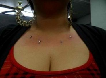
  • On the chest – The chest can be one of the easiest areas to do dermal piercing, but it requires greater caution since the area can be irritated when something like a bath towel accidentally rubs against it.
  • On the back – The lower back, just above the buttocks, can also be a site for piercing. Dermal piercing on the lower back require sleeping on a soft mattress because hard surfaces such as the floor, tough mattresses, and wooden boards may cause friction and pressure on the area.
  • On the neck – The nape area or the side of the neck can be a site for dermal piercing. The skin overlying the collar bone can also be pierced. Piercing the neck area may result in mild swelling and redness because of vast blood supply in the area.
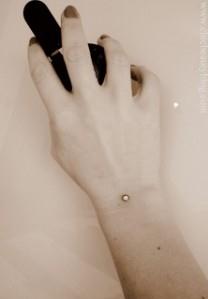
  • On the wrist – Piercing on the wrist is one of the most popular locations for dermal piercing among teens. However, this area is usually not advisable due to the various movements in the wrist and further trauma to the tissues.

Dermal piercing on wrist

Dermal piercing on wrist

  • On the fingers – Piercings can also be placed on one or more fingers to add aesthetic value to the hand.
  • On the face – The face is also one of the most common areas for dermal piercing. It is usually placed on the lips, eyebrows, ears, and tongue.
  • Under the eyes – Dermal piercing under the eyes can be quite complicated and painful, and the procedure should be performed by an experienced professional piercer.

Dermal Piercing Jewelry

Dermal piercing jewelry should be hypoallergenic to reduce irritation and possible rejection. These are most commonly made of metals such as stainless steel, titanium, and niobium.

Among the three, titanium is less likely to cause allergic reactions and irritations. Most jewelry for new piercings which are made from palladium and titanium can be used. Gold and silver are not employed for initial piercings due to possible allergic reactions or metal oxidization (tarnish).

The clasp or stud of the jewelry should also be made with the same material as the primary piece to reduce risk of allergies.

Pictures of Dermal Piercing

Dermal piercing over chest

Dermal piercing on chest

Dermal Piercing Abdomen

Dermal piercing on hips

Neck Piercing

Dermal piercing on neck

Procedure for Dermal Piercing

The procedure for dermal piercing is usually simple but requires precision and care. The step-by-step procedures include:

  1. Disinfecting the piercing site. An antiseptic, usually alcohol, is used to clean the site to remove microorganisms which can potentially cause infection. Hairy sites like the chest and wrist may be shaved for easier procedure.
  2. Marking the site. The area to be perforated is marked with a surgical pen to indicate the entry and exit points of the needle.
  3. Boring the skin. A surgical needle which is sterilized prior to its use is inserted through the skin on the indicated markings.
  4. Inserting the microdermal anchor. The dermal piercing anchor is then placed in the area. When the tissue heals, it usually encapsulates the inserted dermal anchor, making it impossible to remove the anchor without undergoing a surgical procedure.

The whole process usually requires strict asepsis to prevent infection.

Postoperative Care

Following the dermal piercing procedure, aftercare, which is needed to prevent complications, includes the following:

  • Cleaning the site regularly. The site may be cleaned with a mixture of salt and water. Cotton soaked in the solution is used to wipe the area. Salt prevents the occurrence of infection and may not cause significant stinging compared to alcohol. It is advised to clean the site at least twice a day.
  • Washing hands before and after contact with the site. The hands should be maintained clean in order to prevent dirt and other particles from getting trapped in the pierced site.
  • Using mild soap during bathing. Strong soap concentrations may irritate the area.
  • Increasing intake of protein and vitamin C rich foods. Vitamin C strengthens the collagen matrix, thereby preventing infection. Protein enhances wound repair, thereby quickening the healing process.
  • Rinsing away soaps and shampoos effectively. Soap and shampoo can build up on the site and may cause presence of debris. It is advised to rinse off immediately.
  • Removing crusts from the area. Crust from blood and tissues may form on the pierced site. It is important to remove these to prevent further build-up which may be difficult to remove later on.
  • Avoiding frequent touching, fidgeting, and moving the piercing. Frequent contact with the jewelry causes irritation to the surrounding tissues due to friction.
  • Allowing the site to be exposed in the sun from time to time. Vitamin D from sun exposure in the morning can help promote healing. It also prevents infection from anaerobic bacteria, especially in the hidden areas of the body.

Dangers and Risks of Dermal Piercing

Since body piercing is an invasive procedure, it causes several complications such as:

Allergy

Allergy is a possible complication when non-hypoallergenic jewelry is used. Titanium and nobium are the most common hypoallergenic materials which can be used in dermal piercing.

Infection

Infection from viral, bacterial, and fungal origin is also possible. The most common microorganisms which cause infection are the Staphylococcus aureus, pseudomonas, and streptococcal species. Hepatitis B and C can also be acquired when infected needles and equipment are used.

Scarring

During the healing process, hypertrophy of tissues around the area may cause scarring and keloid formation. Infected piercings are more susceptible for scar tissue formation due to abnormal tissue growth and regeneration.

Pain

Pain is an imminent risk for dermal piercing. The perforation in the skin causes the inflammatory response to be activated. Pain can be managed using anti-inflammatory medications and prevented by infrequent contact with the piercings.

Rejection

The body recognizes the piercing as a foreign body that needs to be removed. As a result, the piercing may cause severe pain and produce discharge. A prolonged inflammatory reaction can also be a sign of rejection. When rejection is observed, removal of the piercing is required.

Dermal Piercing Removal

Removal is warranted when there is rejection of the jewelry or when the person chooses to remove the piercing for some reason. Dermal piercings are permanent because the jewelry is already embedded in the tissues after healing has taken place.

The removal requires another surgical procedure to be done by a medical professional in order to detach the jewelry from the tissues. It should never be removed at home because it may cause bleeding and tissue damage.

Dermal Piercing Costs

The general cost of dermal piercings is $75 to $100 in the United States and 40 to 50 pounds in the UK. The price of piercing does not really differ significantly. However, titanium is more cost-effective because it does not cause allergies and rejection.

Resources:

http://tattoo.about.com/od/piercingsgeneralinfo/ss/dermalanchors.htm

http://answers.yahoo.com/question/index?qid=20090503201008AA2WGvY

No responses yet

Lipoatrophy

Jan 03 2013 Published by under Diseases A-Z

What is Lipoatrophy?

Lipoatrophy, also called lipodystrophy, is a condition which describes a loss of fat tissue in a localized area. Lipoatrophy, which usually occurs following frequent subcutaneous injections, is not a disease itself, but is a result of other conditions which lead to a loss of fat tissue in an area.

Lipoatrophy Pictures

Lipoatrophy in the Right Arm

Lipoatrophy or lipodystophy can be partial, localized, or total.

Localized lipoatrophy

Localized lipodystrophy is a localized adipose tissue loss which involves one or more areas. This is the most common form of lipodystophy seen in patients who receive multiple subcutaneous injections.

lipoatrophy

Partial Lipoatrophy

Partial lipodystropy is a symmetrical loss of fat tissue in the face with similar occurrence on the trunk and arms. Partial lipodystophy is correlated with diabetes, renal disease, immunologic disorders, and hyperlipidemia.

Pics of Lipoatrophy on Face of women

Partial Lipoatrophy on the Face

Total lipoatrophy

Total lipodystrophy, which may be acquired or congenital, involves complete loss of adipose tissue.  Causes include insulin resistance, hyperglycemia, hypermetabolism, and hyperlipidemia.

Localized lipodystophy involves single or multifocal, atrophic, well-demarcated lesions. Localized lipoatrophy is rare;  its occurrence without frequent administration of subcutaneous injections is usually rarer. Lipoatrophy is benign and does not result in cancer. There is a low morbidity and mortality rate, and the severity is associated with the involved organ and the occurrence of comorbid conditions. Lipoatrophy is not associated with specific races, and females are more prone to develop it due to fewer muscles beneath the subcutaneous layer. Depending on the presence of causative factors, it can occur at any age. The main concern for lipodystrophy is its cosmetic implication as the appearance of fat loss may be disturbing and can affect the body image of patients.

Most commonly, lipoatrophy may increase in size and may disappear. There are no surface skin changes that happen from the fat loss, but some may experience hyperpigmentation on the skin.

Symptoms of Lipoatrophy

Lipoatrophy manifests as:

  • Presence of depression in the skin in different area.
  • Single or multifocal depression in the skin
  • Well-demarcated and atrophic characteristics
  • Non-pigmented or pigmented lesions
  • Absence of pain
  • Loss of fat in proximal areas of the body (ex., thighs, buttocks, and upper arms)
  • fat loss in face, breasts, scalp, and distal areas of the body (less frequent)

Causes of Lipoatrophy

The exact cause of most lipoatrophy cases is unknown. Local lipoatrophy is usually a result of intradermal and subcutaneous injections. Due to a recurrent trauma to one area of the body as a result of injection, the macrophages release cytokines which lead to increased catabolism or breakdown of adipose tissues. These cytokines include interleukin-1, tumor necrosis factor, histamine, bradykinins, and prostaglandins. Specific medications that lead to lipoatrophy include:

  • Insulin injections

Patients with diabetes who need daily insulin injections are at risk of lipoatrophy. Animal insulin may also lead to lipoatrophy due to its reaction with the fat cells in the body, although this case is very rare.

  • Human growth hormone injections

People with growth hormone deficiency require the administration of growth hormone every day or every week, depending on the physician’s prescription. One study reported the occurrence of lipoatrophy in 13 patients with 3 patients developing it after the first injection.

  • Heparin

Heparin is also injected subcutaneously and may result in lipoatrophy. Heparin also causes hematoma formation when incorrect injection technique is employed.

  • Other synthetic hormones

Other hormones such as erythropoietin are also required to be injected in scheduled doses. The frequent administration of these may lead to localized lipoatrophy.

  • Glatiramer acetate (copaxone)

This drug is used to treat multiple sclerosis. When given subcutaneously, it may lead to lipopatrophy.

  • Corticosteroids

Corticosteroids are also given parenterally, specifically, the intramuscular route. Injections are more common for patients with severe pain and inflammation who do not respond to oral treatments of anti-inflammatory drugs.

  • Iron Dextran

This is an iron preparation administered through the intramuscular route. This may also lead to skin irritation.

  • IM antibiotics

Antibiotics such as penicillins are also injected intramuscularly.

  • Vaccines

Vaccines in the form of diphtheria, pertussis, and tetanus cause reactions in the injection site, which may possibly lead to  lipoatrophy.

Other medication causes of lipoatrophy include HIV lipoatrophy. Lipoatrophy is a common complication of protease inhibitor administration in HIV patients. These are antiretroviral medications that may lead to central fat accumulation and fat tissue loss in other parts of the body. Lipoatrophy caused by HIV medications are usually generalized or total.

Diseases that cause lipoatrophy include:

  • Nephritis
  • Sjögren syndrome
  • Scleroderma
  • Thyroiditis
  • Pyogenic infections
  • Systemic lupus erythematosus
  • Connective tissue disorders

Lipoatrophy is diagnosed mainly by the physical symptoms and physical examinations. Inflammatory lipoatrophies can be detected by the use of serologic markers to determine any connective tissue disease. Histologic examinations, the trademark diagnostic test for lipopatrophy, may be done to determine any cellular changes and will guide the treatment modalities to be employed. Most often, lipoatrophies are benign conditions. Histologic examinations reveal the presence of lobules of small fat cells embedded in connective tissues. This occurrence is termed as involutional fat. There is also the presence of myxoid stroma, peripheral lobular accentuation, and absence of inflammatory cells.

Treatment of Lipoatrophy

Treatment for lipoatrophy is primarily focused on surgery. There are no medications, diet, and activity that will correct the problem. Fat loss under the skin will not be replaced with certain medical treatments.

Surgery

Surgery is the treatment of choice for lipoatophy. Patients who are very conscious about their appearance, especially when the lipoatrophy occurs on the face, would opt for surgical treatment to regain aesthetic confidence. Lipoatrophy when left untreated with surgery may not cause other complications. Aesthetic reasons are the primary considerations for surgery.

The surgical procedure employed is the placement of adipofascial flaps under the skin to even out the integumentary system and correct skin depressions.

Lipoatrophy Treatment images

Lipoatrophy Surgery involving Placement of Flaps

Alternative treatments

Management for lipoatrophy caused by insulin injections require the shift from animal insulin to purified human insulin.

Prevention of Lipoatrophy

Localized lipoatrophy is primarily prevented by rotation of injection sites. Everyday injections should be administered in different parts of the body to minimize overuse of a certain spot which may cause lipoatrophy. Other conditions leading to lipoatrophy should also be managed to prevent the loss of fat tissue.

Prognosis of Lipoatrophy

Patients with lipoatrophy have a normal life expectancy and will not experience other complications aside from cosmetic affectation. Lipoatrophy is usually benign.

No responses yet

© 2017 HealthZene.com. All Rights Reserved. Privacy Policy
This website is for informational purposes only and Is not a substitute for medical advice, diagnosis or treatment.