Hereditary Hemorrhagic Telangiectasia – Treatment, Diagnosis, Symptoms
What is Hereditary Hemorrhagic Telangiectasia?
Contents
Hereditary hemorrhagic telangiectasia (HHT) is a genetic condition that causes the abnormal formation of blood vessels in the mucous membranes, brain, liver, lungs, and skin. The presence of abnormal blood vessel formation may lead to gastrointestinal bleeding, nose bleed, and other organ problems.
Symptoms of Hereditary Hemorrhagic Telangiectasia
Symptoms of HHT are related to abnormal blood vessels that may lead to easy bleeding. Symptoms include:
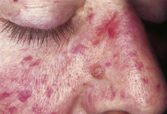
Small vascular formations in the skin and mucous membranes
The small, thin vascular malformations are called telangiectasias which may be apparent on the skin of the face, legs, mouth, lips, fingers, tongue, and other sun-exposed areas. The lesions may bleed, but the most common concern is that these small vessels are cosmetically displeasing. The telangiectasias often appear suddenly and increase in number over time.
Arteriovenous malformation (AVM)
These are larger vascular malformations that occur in major organs of the body such as the brain, liver, lungs, and even the spinal cord. Blood clots in the brain may lead to stroke, and the inability to filter out bacteria may lead to brain abscess when the infection reaches the brain. Bleeding in the lungs may cause hemoptysis (coughing up blood).
Breathlessness
Breathlessness is also a sign of AVM in the lungs. AVM allows the unoxygenated blood to bypass the alveoli; thus there is poor blood oxygenation which causes breathlessness. There may also be breathing difficulty which is more severe when sitting than lying down.
Cyanosis
Poor blood oxygenation in the lungs may also lead to cyanosis due to severe hypoxia. Chronic hypoxia may also lead to clubbing of the fingers.
Epistaxis
Epistaxis or nose bleeding is a common problem associated with HHT. Nose bleed is a common occurrence in childhood and may affect up to 95 percent of people with HHT. Nose bleed results from the rupture of the telangiectasia found on the mucosal lining of the nasal cavity.
Blood in vomitus
Blood appearing in the vomitus may indicate bleeding in the stomach or esophagus due to lesions in the upper gastrointestinal tract.
Black stool
Black stool, which may also be apparent, is a sign of bleeding of small lesions in the lower gastrointestinal tract.
Anemia
Anemia may be due to various small bleeds in the body such as in the gastrointestinal tract (GIT) and nose. Patients may experience iron deficiencythat may manifest as paleness and fatigue.
Congestive heart failure may develop when the heart compensates for the limited amount of blood that goes into the organs due to large vascular malformations. Affectation of the liver may cause portal hypertension and eventually ascites (the presence of accumulated fluid in the peritoneal cavity). Portal hypertension may also lead to esophageal varices.
Genetics and Pathophysiology
HHT is an autosomal dominant trait, i.e., the offspring may have a 50-percent chance of acquiring the disorder. However, new mutations have been documented in people with no HHT in the family. Affected people carry one abnormal gene of HHT. It appears that carrying two abnormal copies of the gene is not compatible with life); thus no homozygotes have been documented. Genetic mutations in HHT are often associated with mutations in ACVRL1 or ENG gene. There are a total of 600 gene mutations that were known to cause HHT.
The pathophysiological mechanism of HHT involves changes in angiogenesis (development of blood vessels). Angiogenesis requires the activation of smooth muscles, endothelium, and pericytes. In HHT, however, there is a disruption in the balance on the pro- and antiangiogenic signals in the vasculature, thereby leading to friable growth of blood vessels. Being friable, the vasculature has an increased tendency to bleed.
Diagnosis
There are many diagnostic tests for HHT which are used to confirm the presence or absence of HHT, and to identify complications of HHT. Diagnostic tests include:
- Physical examination
Physical examination usually identifies telangiectasia in the skin and mucous membranes. Laryngoscopy and endoscopy are done to detect lesions in the nose and larynx.
- Esophagogastroduodenoscopy (EGD)
Esophagogastroduodenoscopy usually detects the presence of telangiectasia in the digestive tract through direct visualization of the gastrointestinal tract (GIT) mucosa. Capsule endoscopy may be employed to detect telangiectasia in the small intestines.
- Chest X-ray or radiology
Chest X-rays are used to identify the presence of AVM in the lungs. Additional pulse oxymetry may be used to identify hypoxia caused by AVM in the lungs.
- Bubble contract echocardiography
This test is used to detect abnormal connections in the arteries and veins in the lungs. The lungs normally remove air bubbles from the circulation. The presence of air bubbles in the left chambers of the heart may indicate lung or heart AVMs.
- CT scan
CT scan may be done to detect lung lesions. It is also used to detect AVMs in other organs such as the brain.
- Cardiac catheterization
This is often employed to determine the presence of increased pressure in the right chambers of the heart due to liver congestion.
- Doppler ultrasonography
This test is used to detect problems in the liver such as the presence of vascular lesions.
- Genetic testing
Genetic testing is not often employed to ascertain the diagnosis, but it is used to identify the specific gene mutation that may have caused the disorder.
Criteria for HHT
In order to diagnose HHT, Curacao criteria is employed. The presence of three or all of the four criteria indicates the definite presence of HHT, while that of two of the four criteria indicates a possible presence of HHT.
- Recurrent and spontaneous epistaxis
- HHT in first-degree family member
- Presence of multiple telangiectasia in typical locations
- Presence of visceral AVM (in the spine, brain, liver or lungs)
Treatment of Hereditary Hemorrhagic Telangiectasia
Treatment regimens for HHT include intervention aimed at reducing the bleeding from the vascular malformations and other supportive treatments. These include:
Iron supplementation
Chronic and recurrent bleeding may lead to anemia; thereby iron supplementation is important to prevent iron deficiency states.
Blood transfusion
Blood transfusion is also employed in order to correct hypovolemia states or (reduced blood components) due to heavy bleeding.
Managements for nose bleed
Atraumatic nasal packing
Nasal packing may be placed for patients who experience acute nose bleed. Preventing nose bleed involves keeping the nasal cavity moist with the use of saline solution.
Nasal coagulation and cauterization
These modalities are used to prevent nose bleed from telangiectasia.
Interventional radiology
This measure involves the embolization of vascular lesions to prevent bleeding. The procedure involves the passing of a thin catheter through the arteries and the injection of substances to occlude the blood vessels. This procedure is appropriate for severe bleeding.
Surgery
Surgery becomes the last resort for HHT treatment. Saunder’s procedure or septal dermoplasty involves transplantation of skin in the nostrils. Young’s procedure, which involves the permanent closure of the nostrils may also be done.
Managements for organ AVM
The presence of organ AVMs is managed by using embolization to prevent organ bleeding. Metal coils may also be placed. AVMs in the brain may require craniotomy to locate the vascular malformations.
Pictures